The science of sea urchins
Greenhills anatomy and physiology students had the exciting opportunity last week to view fertilization first hand using gametes harvested from live sea urchins.
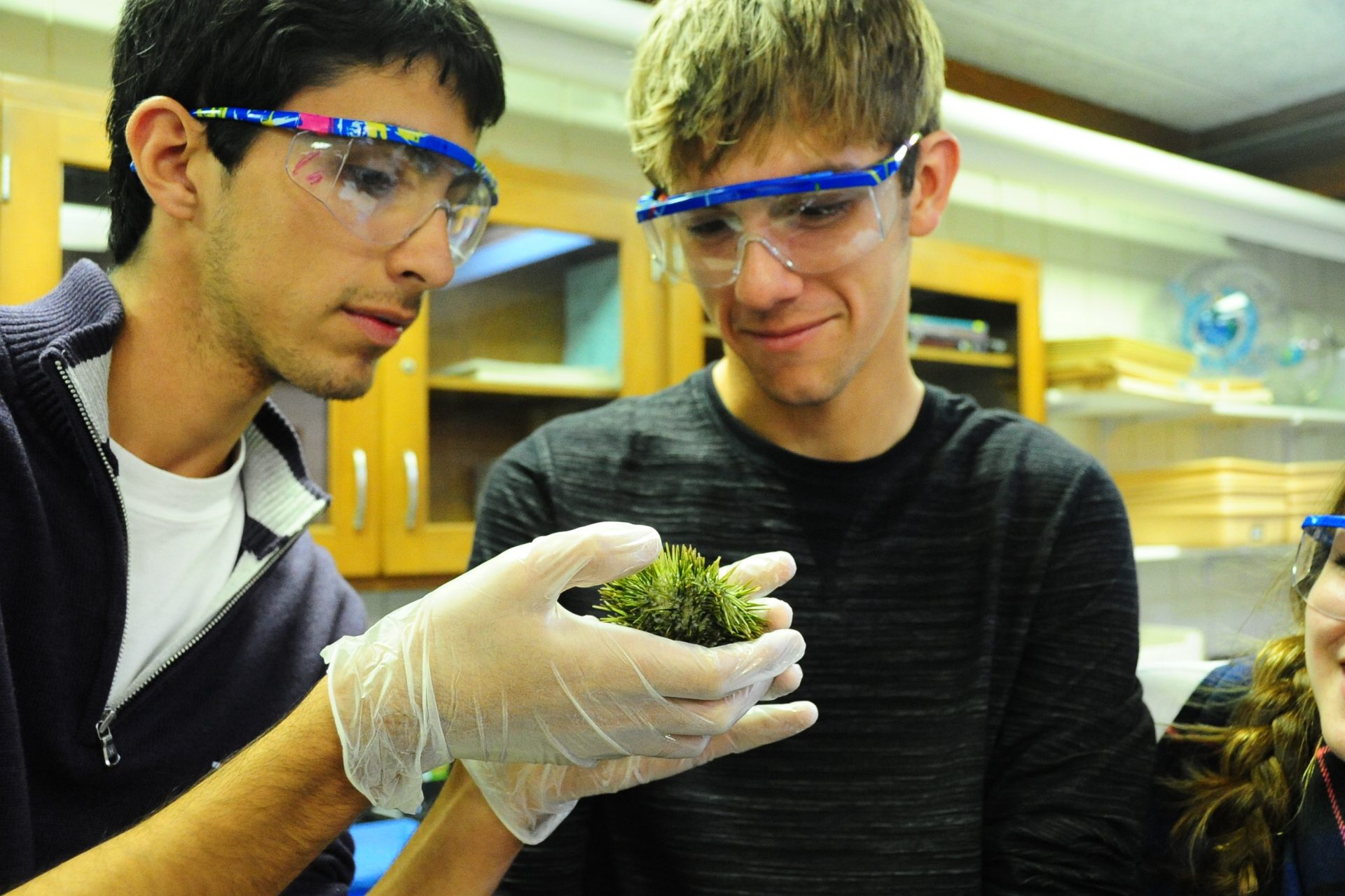
 The sea urchins traveled all the way from the Florida coast, bringing with them a salty sea smell and lots of opportunity for discovery. Anatomy students exclaimed, “How cute,” and “Look at its mouth,” as they gently handled the living urchins. Students had explored several websites including Stanford University’s virtual urchin site, in preparation for this week’s visit.
For the second part of the lab, students watched as I injected four urchins with a solution of potassium chloride. This solution caused the urchins to begin producing either eggs or sperm. There is no way to identify whether the urchin is male or female until you see these cells.
Once the cells were released and identified, students took samples and observed them separately using a microscope. Students were shocked at how small and numerous the sperm were, finding them best under 400x magnification. They were equally surprised by the fact that they could see the urchin eggs with their naked eyes.
Then, the students added one drop of the dilute sperm solution to their egg slide and observed the changes. Students noted an almost immediate change to the cell membrane of the egg, and later some even observed cell division.
The urchins were frozen overnight, then thawed the next day to allow students to dissect them. Students were especially impressed by the structure of the five-part teeth, termed Aristotle’s lantern.
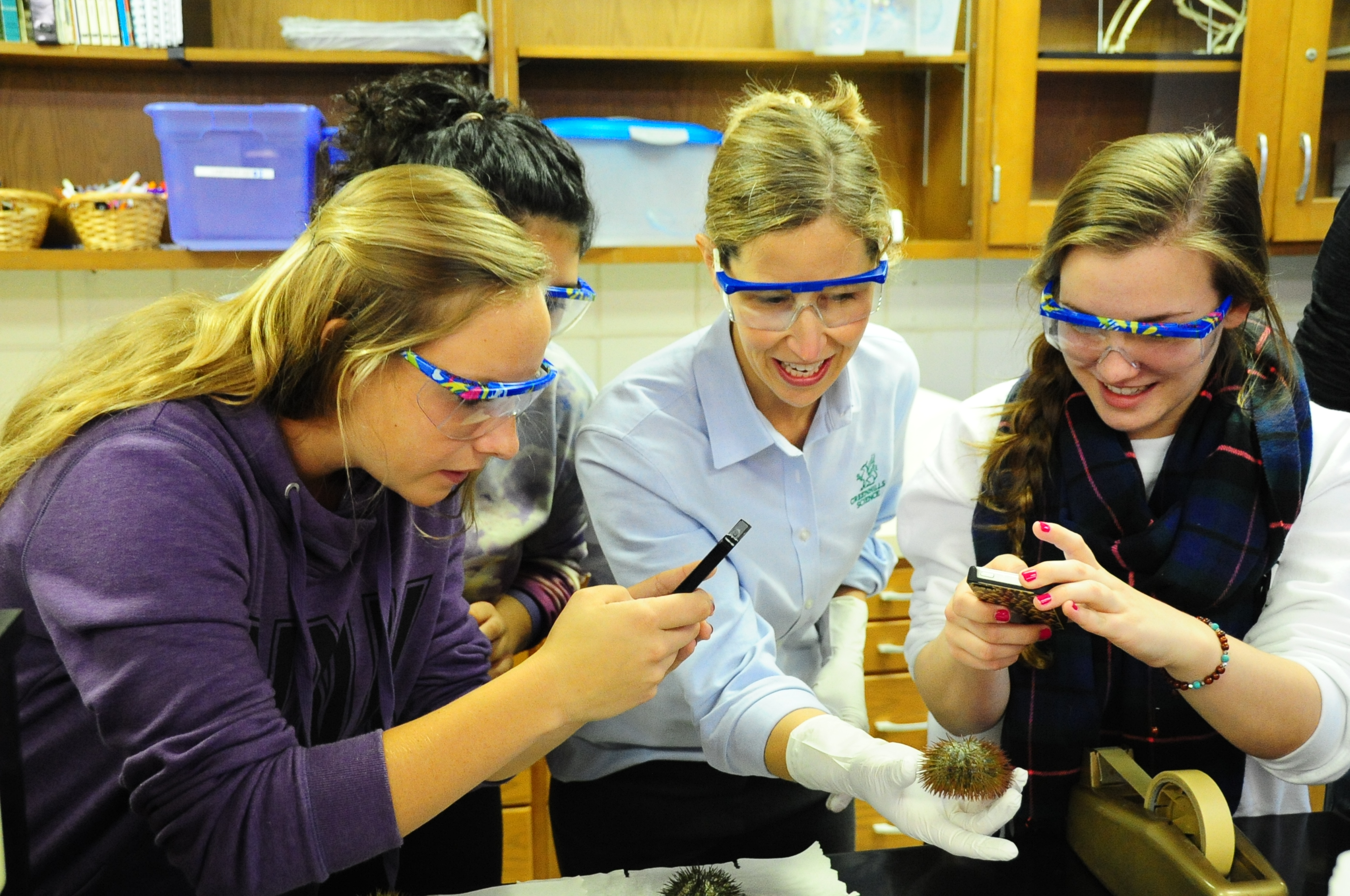
Using cameras on their cell phones, students documented the dissection, noting interesting structures and talking about how gross the digestive tract looked.
One junior, who had admitted to eating live urchins this past summer, exclaimed, “I can’t believe I ate this stuff.”
–By Dee Lamphear, Greenhills science teacher


